Experience Opportunities Research Symposium

The 29th Annual Westmont College Spring Student Research Symposium
Thursday, April 17th, 2025 from 3:30-5:00PM
In the Winter Hall Atrium
One of the hallmarks of Westmont College's academic program is the opportunity for undergraduate students to work directly with faculty on research projects. Work presented at the annual Spring Student Research Symposium includes student research conducted during the past year from all three divisions of the college (Humanities, Social Sciences, and the Natural and Behavioral Sciences).
We look forward to soaking in the original research of our students and celebrating their tremendous accomplishments!
Click Here to See What Types of Research Students Are Working On
Who should apply?
Any student in any major who has or is participating in a semester-long or longer research project under the supervision of a faculty member during this academic year can apply. This would include, but is not limited to, major honors students, independent research students, and students in a class with a significant cumulative research project (e.g. some senior capstone classes). Students working in partnership on the same project should apply together.
Symposium format
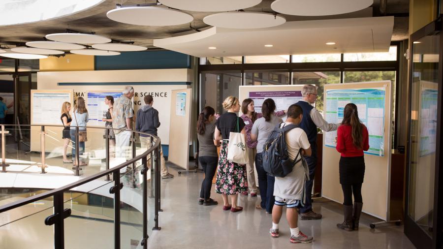
Each presenter or team prepares an overview of their research project in the form of a poster (max size: 48" x 36"). All posters are displayed throughout the 1 1/2-hour event and presenters stand by their poster to discuss the research with visitors and answer questions.
Poster design and printing
Posters are usually made as one large “slide” in PowerPoint or Keynote, or in another graphics program. Printing can be done either locally and picked up or online and shipped here. Some options are below. In designing posters, students should keep in mind the physical distancing requirements and that attendees will not be able to get within 6 feet of the poster. We suggest less text, larger font for the text that is included, and more visuals like schematics, pictures, graphs, and tables.
Poster Nerd Remote, but ships quickly. (also has templates you can download into PowerPoint)
Tri-Co Reprographics at 720 E. Haley St.
Bill's Copy Shop at 1536 State St.
Westmont Reprographics (may have limited availability and would need a minimum of 3 days lead time; 24” max width)
How to apply
Complete the online abstract submission form. Use the sample, below, for guidance of how it should be formatted.
DEADLINE IS MONDAY, March 31st, 2025
Sample title/author list/abstract
The Role of TDP-43 Oligomerization of ALS Neurodegeneration
Brady Quon (’16) and Megan Korff (’16), Faculty Mentor: Kristi Cantrell, Chemistry
Author Email(s): author1@westmont.edu, author2@westmont.edu
Abstract : TAR-DNA-binding protein (TDP-43) is an RNA processing protein that is primarily located in the nucleus. Recent research supports that TDP-43 aggregation into fibrils and oligomers, as well as translocation into the cytoplasm, are characteristic of the neurodegenerative disease Amyotrophic Lateral Sclerosis (ALS). Several ALS-associated mutations have been found in the carboxyl terminal, glycine rich region of TDP-43. Six, 13 residue peptide fragments are currently being synthesized to elucidate the conformation and aggregation of the TDP-43 glycine-rich region. Fibril assays have been prepared from two of the purified peptides and imaged using electron microscopy. Thioflavin T was applied to any fibrillar aggregates to determine the presence of beta-sheet secondary structure.